kilasan memoir
Sunday, February 8, 2009
cells biology
Cell biology (also called cellular biology or formerly cytology, from the Greek kytos, "container") is an academic discipline that studies cells – their physiological properties, their structure, the organelles they contain, interactions with their environment, their life cycle, division and death. This is done both on a microscopic and molecular level. Cell biology research encompasses both the great diversity of single-celled organisms like bacteria and protozoa, as well as the many specialized cells in multicellular organisms like humans.
Knowing the components of cells and how cells work is fundamental to all biological sciences. Appreciating the similarities and differences between cell types is particularly important to the fields of cell and molecular biology as well as to biomedical fields such as cancer research and developmental biology. These fundamental similarities and differences provide a unifying theme, sometimes allowing the principles learned from studying one cell type to be extrapolated and generalized to other cell types. Hence, research in cell biology is closely related to genetics, biochemistry, molecular biology and developmental biology.
Contents |
Processes
Movement of proteins
Each type of protein is usually sent to a particular part of the cell. An important part of cell biology is the investigation of molecular mechanisms by which proteins are moved to different places inside cells or secreted from cells.
Most proteins are synthesized by ribosomes in the cytoplasm. This process is also known as protein biosynthesis or simply protein translation. Some proteins, such as those to be incorporated in membranes (known as membrane proteins), are transported into the endoplasmic reticulum (ER) during synthesis. This process can be followed by transportation and processing in the Golgi apparatus. From the Golgi, membrane proteins can move to the plasma membrane, to other subcellular compartments, or they can be secreted from the cell. The ER and Golgi can be thought of as the "membrane protein synthesis compartment" and the "membrane protein processing compartment", respectively. There is a semi-constant flux of proteins through these compartments. ER and Golgi-resident proteins associate with other proteins but remain in their respective compartments. Other proteins "flow" through the ER and Golgi to the plasma membrane. Motor proteins transport membrane protein-containing vesicles along cytoskeletal tracks to distant parts of cells such as axon terminals.
Some proteins that are made in the cytoplasm contain structural features that target them for transport into mitochondria or the nucleus. Some mitochondrial proteins are made inside mitochondria and are coded for by mitochondrial DNA. In plants, chloroplasts also make some cell proteins.
Extracellular and cell surface proteins destined to be degraded can move back into intracellular compartments upon being incorporated into endocytosed vesicles. Some of these vesicles fuse with lysosomes where the proteins are broken down to their individual amino acids. The degradation of some membrane proteins begins while still at the cell surface when they are cleaved by secretases. Proteins that function in the cytoplasm are often degraded by proteasomes.
Other cellular processes
- Cell division - a eukaryotic cell process resulting in the formation of daughter cells; there are two major types, that of mitosis and meiosis, asexual reproduction and sexual reproduction, respectively.
- Cell signaling - Regulation of cell behavior by signals from outside.
- Active transport and Passive transport - Movement of molecules into and out of cells.
- Adhesion - Holding together cells and tissues.
- Transcription and mRNA splicing - gene expression.
- Cell movement: Chemotaxis, Contraction, cilia and flagella
- DNA repair and Cell death
- Metabolism: Glycolysis, respiration, Photosynthesis
- Autophagy - The process whereby cells "eat" their own internal components or microbial invaders
Internal cellular structures
- Organelle - term used for major subcellular structures
- Chloroplast - key organelle for photosynthesis
- Cilia - motile microtubule-containing structures of eukaryotes
- Cytoplasm - contents of the main fluid-filled space inside cells
- Cytoskeleton - protein filaments inside cells
- Ribosome - RNA and protein complex required for protein synthesis in cells
- Endoplasmic reticulum - major site of membrane protein synthesis
- Flagella - motile structures of bacteria, archaea and eukaryotes
- Golgi apparatus - site of protein glycosylation in the endomembrane system
- Membrane lipid and protein barrier
- Lipid bilayer - fundamental organizational structure of cell membranes
- Vesicle - small membrane-bounded spheres inside cells
- Mitochondrion - major energy-producing organelle by releasing it in the form of ATP
- Nucleus - holds most of the DNA of eukaryotic cells and controls all cellular activities
Techniques used to study cells
Cells may be observed under the microscope. This includes the Optical Microscope, Transmission Electron Microscope, Scanning Electron Microscope, Fluorescence Microscope, and by Confocal Microscopy.
Several different techniques exist to study cells.
- Cell culture is the basic technique of growing cells in a laboratory independent of an organism.
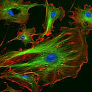
cells as they divide and migrate ken
- Immunostaining, also known as immunohistochemistry, is a specialized histological method used to localize proteins in cells or tissue slices. Unlike regular histology, which uses stains to identify cells, cellular components or protein classes, immunostaining requires the reaction of an antibody directed against the protein of interest with the tissue or cell. Through the use of proper controls and published protocols (need to add reference links here), specificity of the antibody-antigen reaction can be achieved. Once this complex is formed, it is identified via either a "tag" attached directly to the antibody, or added in an additional technical step. Commonly used "tags" include fluorophores or enzymes. In the case of the former, detection of the location of the "immuno-stained" protein occurs via fluorescence microscopy. With an enzymatic tag, such as horse radish peroxidase, a chemical reaciton is carried out that results in a dark color in the location of the protein of interest. This darkened pattern is then detected using light microscopy.
- Gene knockdown mutates a selected gene
- Transfection introduces a new gene into a cell, usually an expression construct
- PCR can be used to determine how many copies of a gene are present in a cell
- In situ hybridization shows which cells are expressing a particular RNA transcript
- DNA microarrays identify changes in transcript levels between different experimental conditions
- Computational genomics is used to find patterns in genomic information [1]
Purification of cells and their parts
Purification may be performed using the following methods:
- Flow cytometry
- Cell fractionation
- Release of cellular organelles by disruption of cells.
- Separation of different organelles by centrifugation.
- Proteins extracted from cell membranes by detergents and salts or other kinds of chemicals.
- Immunoprecipitation.


0 Comments:
Post a Comment
<< Home